Last updated April 27, 2018 at 10:40 am
Telomerase may not have delivered the fountain of youth but it still holds immense promise.
When Australian scientist Elizabeth Blackburn announced the discovery of telomerase – the enzyme that protects the ends of chromosomes and prevents them from fraying enough to kill a cell – there was widespread hope it could lead to the discovery of a drug to slow ageing, and a new breed of anticancer therapies.
Thirty years on and there are still no telomerase-based anti-ageing drugs touted as a “fountain of youth,” nor anticancer drugs.
However, we may have moved one big step closer with researchers from UC Berkeley revealing the detailed structure of human telomerase for the first time.
Like someone removing a blindfold from an archer to reveal the target, scientists finally have a finely detailed 3D structure of the enzyme which they can use to design highly specific and effective drugs.
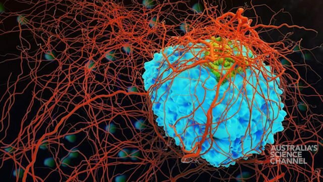
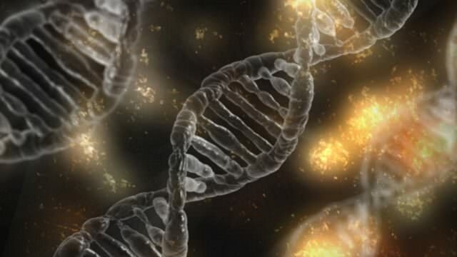
A human chromosome with telomeres highlighted. Credit: AJC1/Flickr
The chromosome’s protector
Telomeres are protective caps found at the end of chromosomes – the twisted and packed structures of DNA inside our cells.
In a way, they’re similar to aglets (the plastic tips found on shoelaces) because they prevent the ends of the chromosomes from ‘fraying.’
However, every time a cell divides, the telomeres become shorter and shorter until eventually the cell stops dividing and dies.
Telomerase is the telomere repair enzyme, adding extra DNA to the telomeres to increase their length and prevent the cell from reaching the point where it dies.
This has two effects. Telomerase could lengthen the life of cells, allowing more divisions before it dies and thereby slow or halt the ageing process. But if this becomes out of control – a cell that can endlessly replicate without dying can be the seed for cancer.
Indeed, the majority of cancerous tumours have activate telomerase doing exactly that, while normal human cells have inactive telomerase (with a few exceptions such as sperm, developing embryos, and some immune cells).
As active telomerase gives cells the potential to become tumours, and that elevated telomerase activity is found in cancer cells, cancer researchers have long searched for a drug which will inhibit the enzyme.
Preventing this activity would reverse cancer growth, essentially making the tumour kill itself from old age, while leaving normal human cells (with their already inactive telomerase) relatively untouched.
While there are some telomerase inhibitor molecules available, their clinical usefulness is limited as they require quite high doses. The new structure, however, could allow brand new studies using computer simulations to discover likely drug binding sites on telomerase and drugs which may be able to interact with them. All of a sudden, drug discovery can be a lot more strategic.
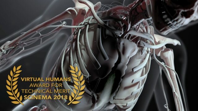
The structure of the active human telomerase. Schematic of subunit arrangements (left), and Front (middle) and
back (right) views of the enzyme. Credit: Nguyen et al, Nature
Telomerase’s structure
Telomerase has an exceptionally complex structure, with an RNA backbone decorated by six types of protein that move around as they add DNA to the ends of chromosomes.
With the complex structure comes complex questions, such as whether the enzyme operates singly or as conjoined twins, and how and just how many proteins decorate the RNA backbone.
Without answers to these questions, it has proven difficult to design a drug to target the molecular machine and either destroy telomerase activity, or indeed restart telomerase, such as to boost cell division after a bone marrow transplant.
The Berkeley lab has been trying to determine the structure of telomerase for 20 years and had made some advances before now, including discovering and characterising many of the proteins in the enzyme, as well as the broken-up hairpin structure of the RNA backbone of telomerase.
However, they still weren’t sure how they all fit together because of conflicting results from many different labs.
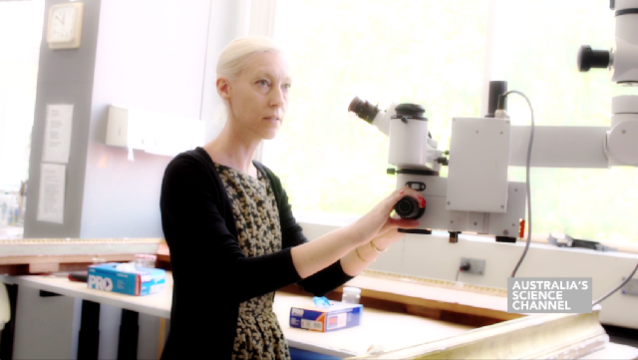
The breakthrough came when lab member Thi Hoang Duong “Kelly” Nguyen was able to isolate the active enzyme and purify it much better than anyone had before. From there, they used a new, state-of-the-art microscope called a cryoelectron microscope.
The cryoelectron microscope, which fires beams of electrons at a sample of frozen proteins, allowed them to determine the structure of the active telomerase unambiguously.
“When I got to the point where I could see all the subunits – we had 11 protein subunits in total – it was a moment of, ‘Wow, wow, this is how they all fit together,'” said Nguyen.
The head of the laboratory Kathleen Collins, who has devoted much of her career to finding telomerase’s structure, said that the years of work had been worth it. “I didn’t think it would be this complicated when I decided to study this molecule,” she said. “It has been a long time coming. It took a lot of persistence.”
While it is the highest resolution image of telomerase ever produced – increasing the resolution about four-fold, the newly revealed structure still lacks fine detail, but combined with knowledge of the gene sequence of human telomerase, it provides enough information to start thinking about potential targets for drugs.
The researchers are now working to improve the resolution even further to about the size of two carbon atoms – which will unlock all sorts of possibilities when it comes to drug design.
The research has been published in Nature
Research video courtesy of Thi Hoang Duong Nguyen, Jane Tam, Robert A. Wu, Basil J. Greber, Daniel Toso, Eva Nogales, Kathleen Collins and InMotionScience